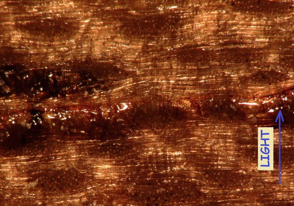
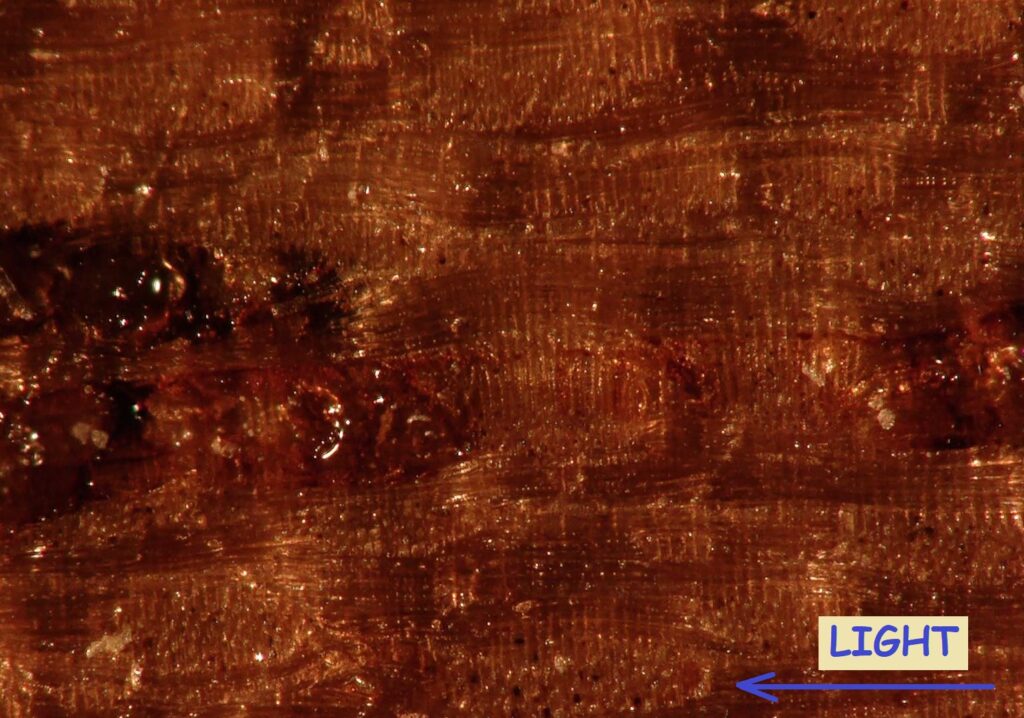
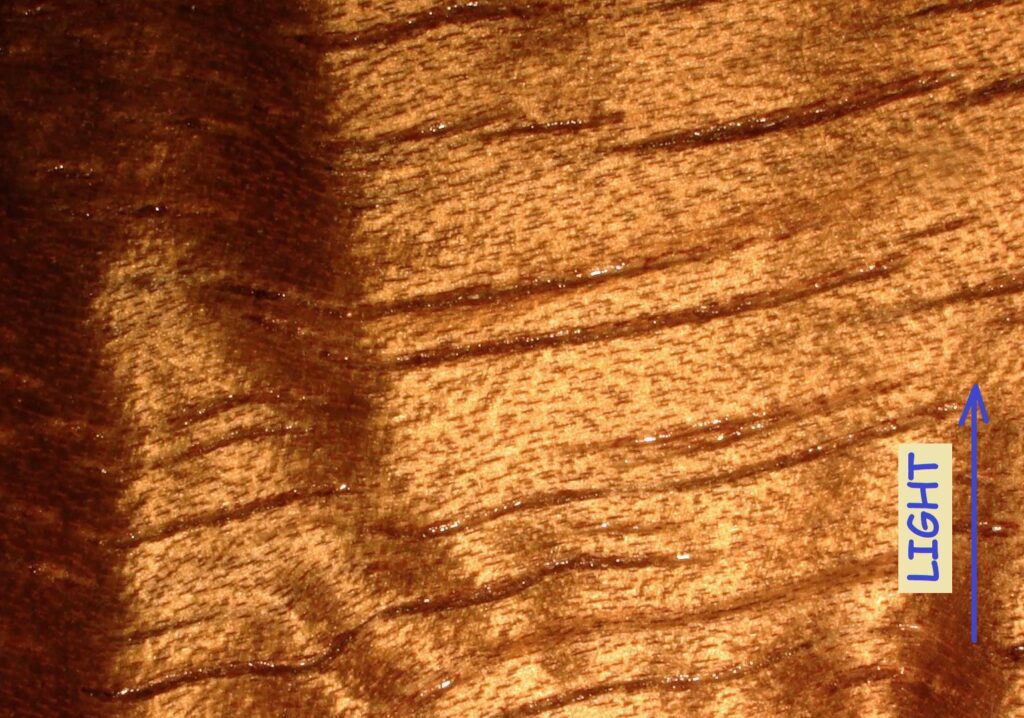
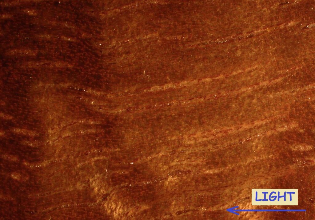
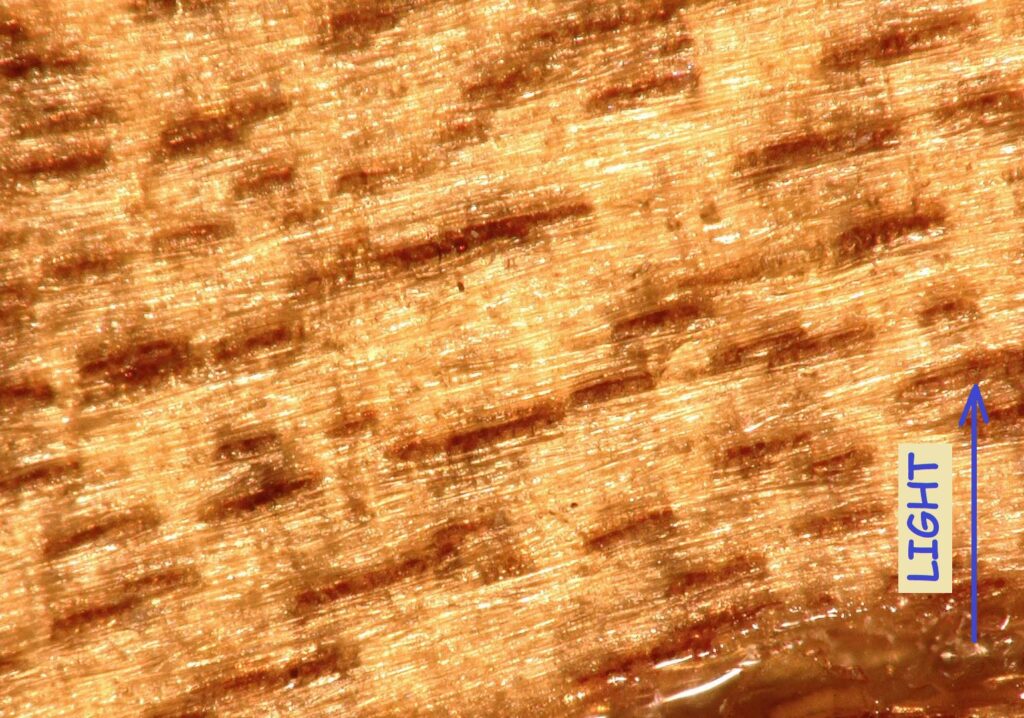
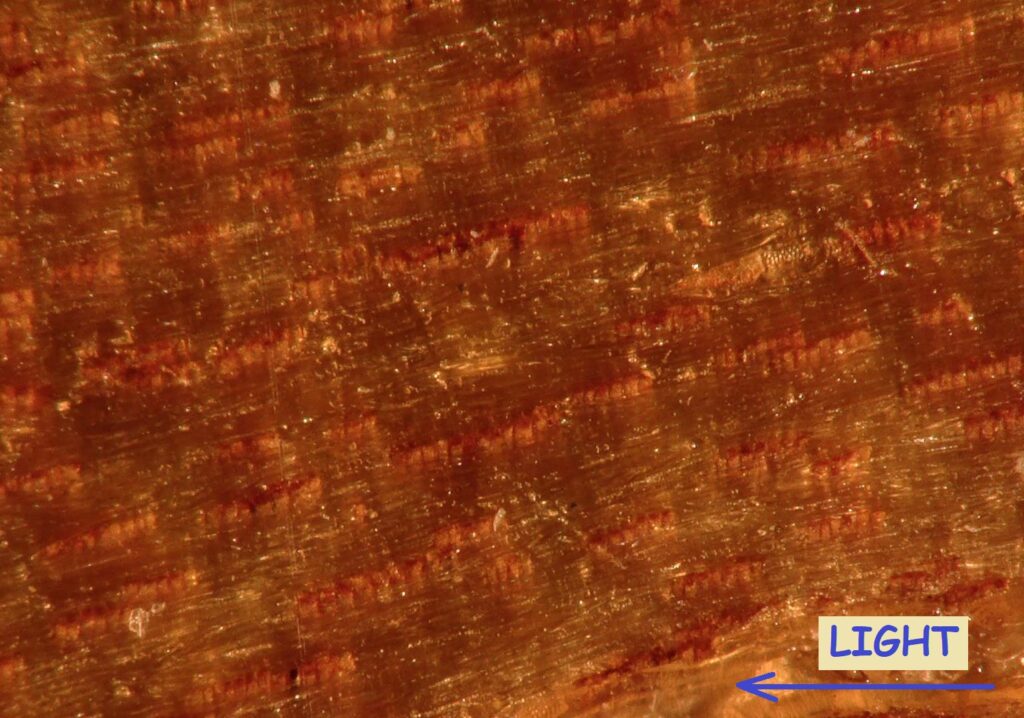
Seven samples were sanded up to 10’000-grit, coated with shellac and then inspected through microscope under two different magnification levels, and each was inspected under two different lighting directions. Koa provided the clearest view of the difference caused by different lighting directions, summarized in the following 1mm x 1mm pictures:
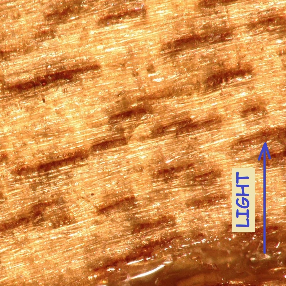
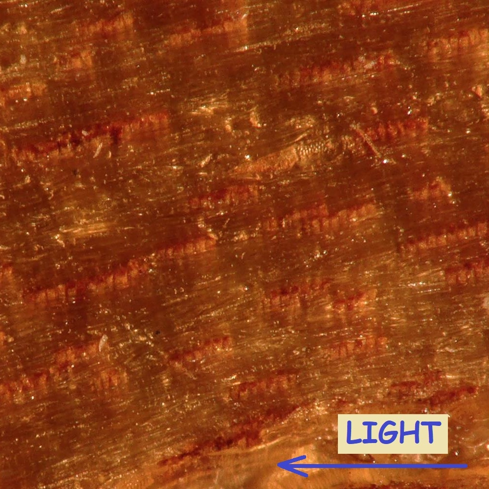
Under this magnification it appears that the cause of chatoyance are fibers themselves; however, a much higher magnification is required to better understand the phenomenon.
All resulting pictures are reported here:
Spanish Cedar (pictures representing a 15x11mm area):
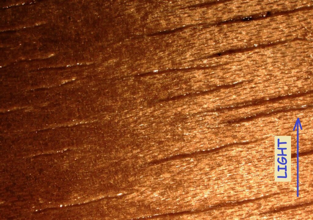
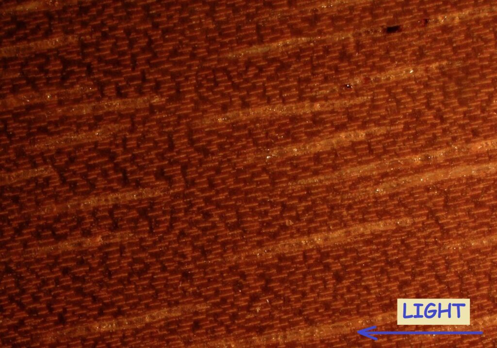
Spanish Cedar (pictures representing a 1.5×1.1mm area):
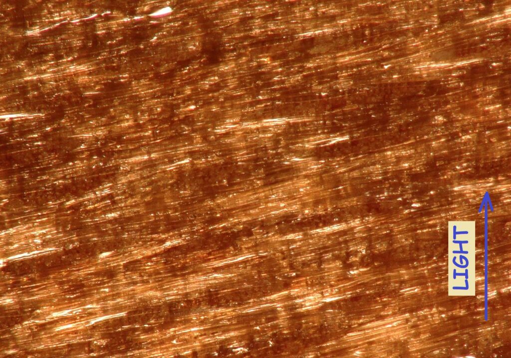
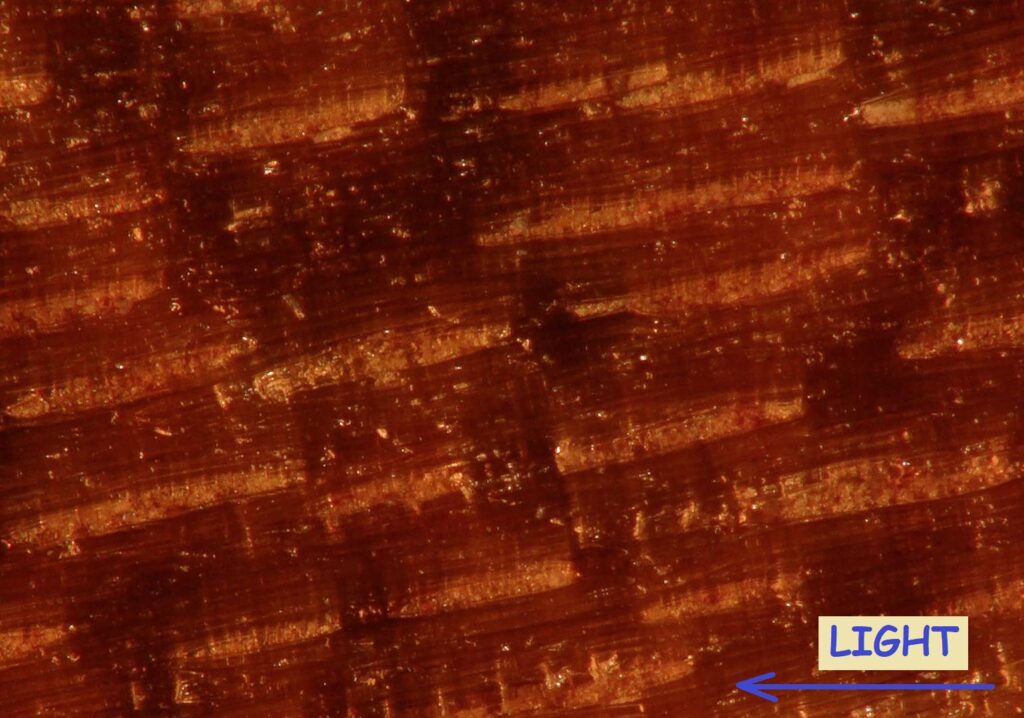
Iroko (pictures representing a 15x11mm area):
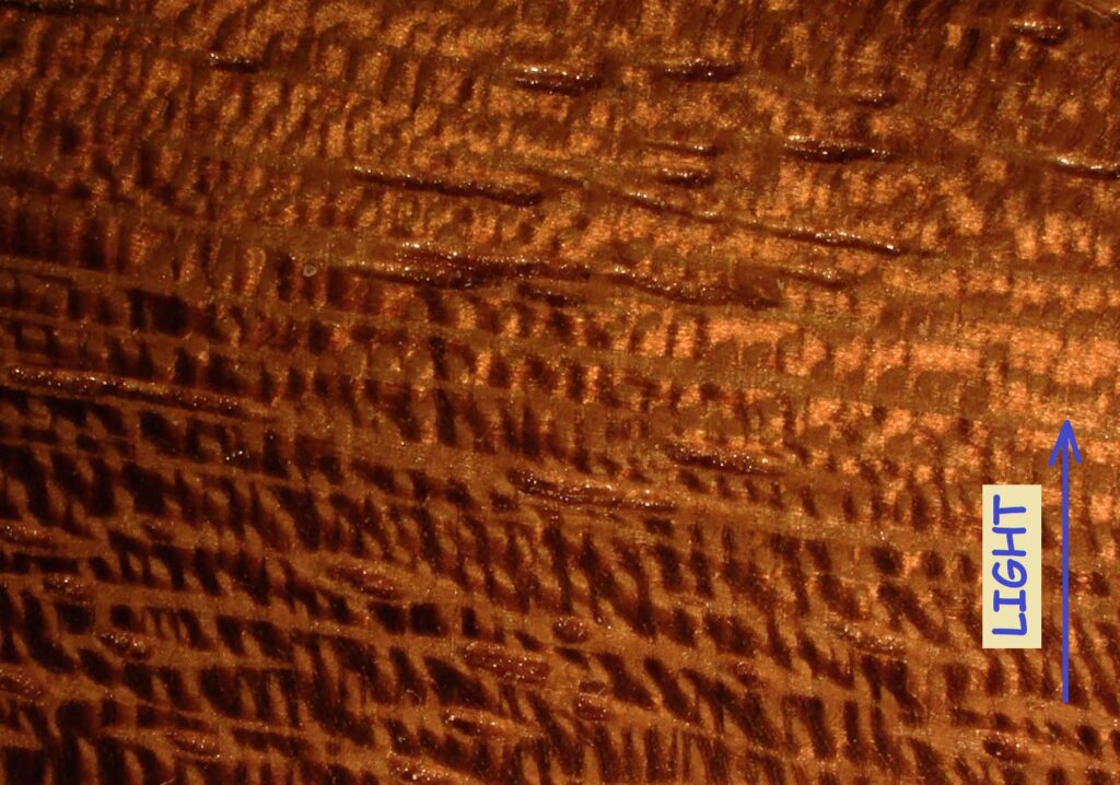
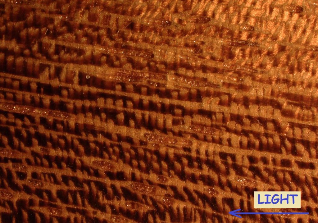
Iroko (pictures representing a 1.5×1.1mm area):
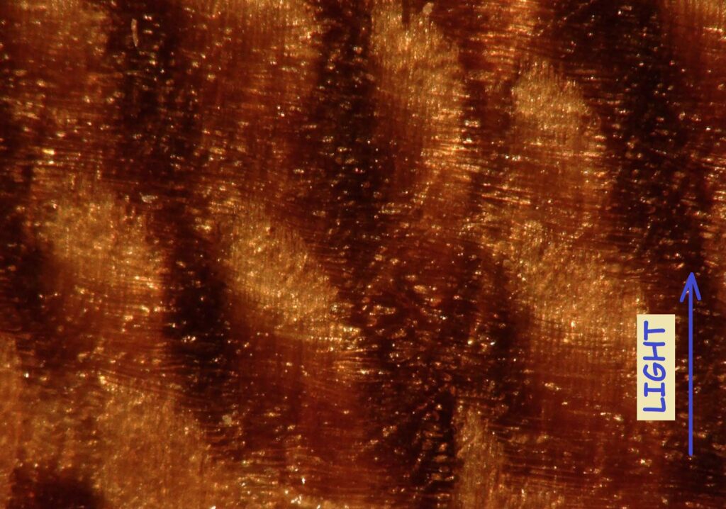
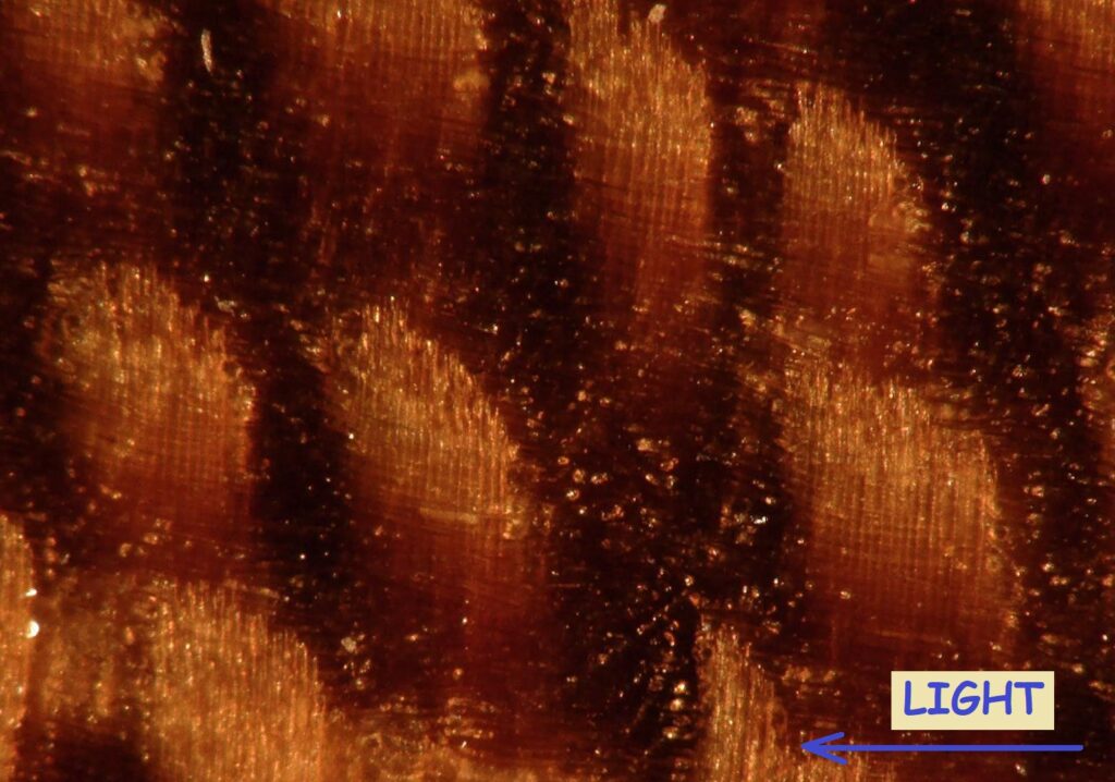
Koa (pictures representing a 15x11mm area):
Koa (pictures representing a 1.5×1.1mm area):
Populus spp. (pictures representing a 15x11mm area):
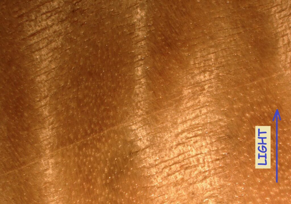
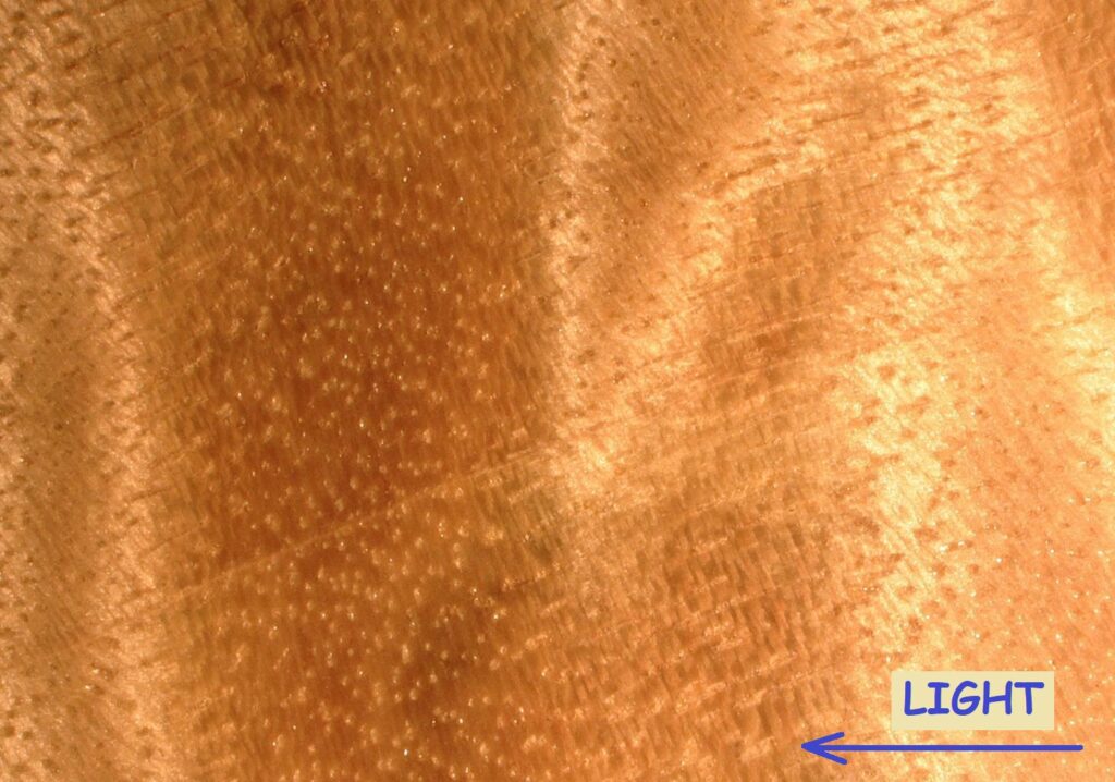
Populus spp. (pictures representing a 1.5×1.1mm area):
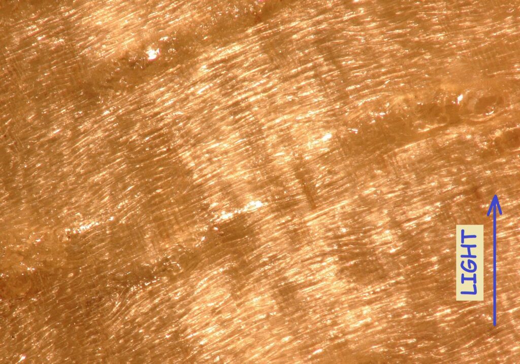
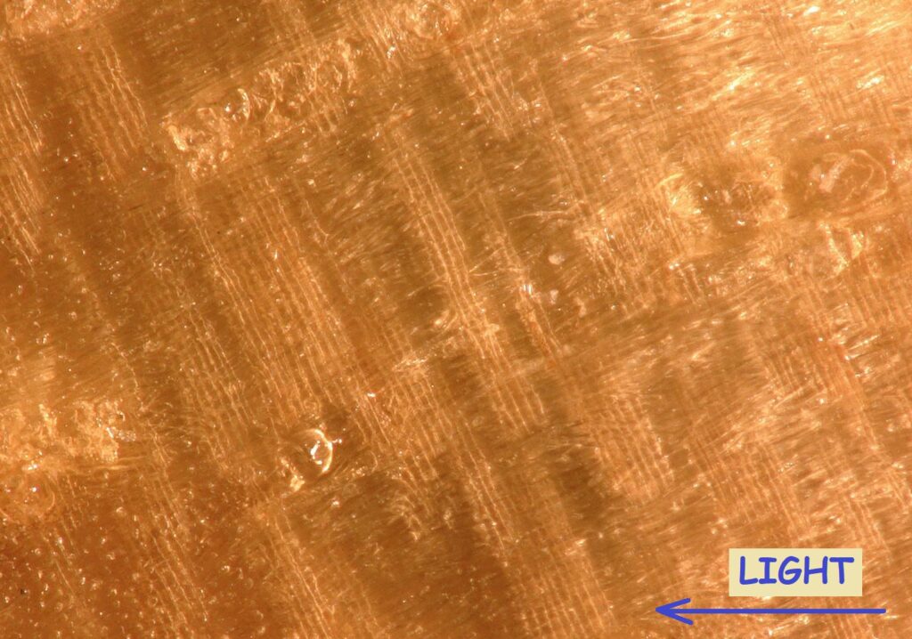
Black Locust (pictures representing a 15x11mm area):
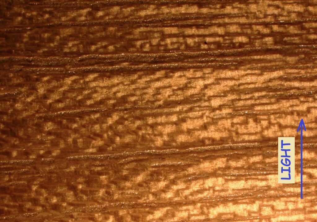
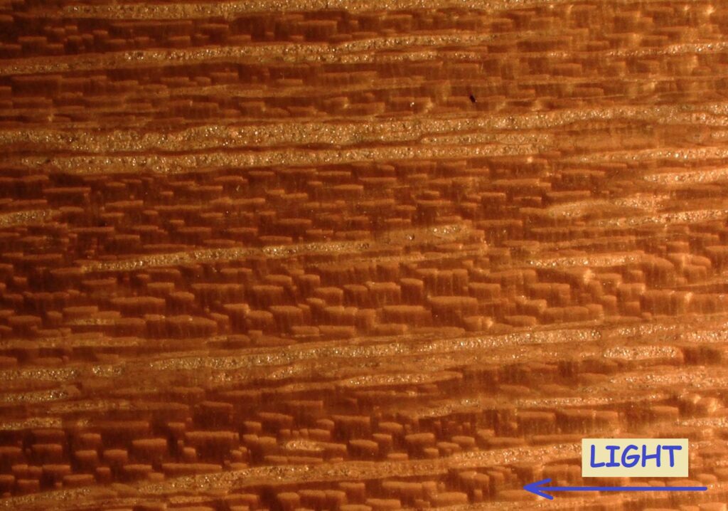
Black Locust (pictures representing a 1.5×1.1mm area):
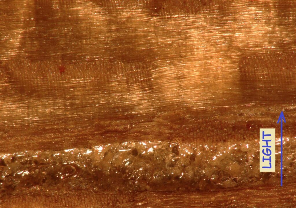
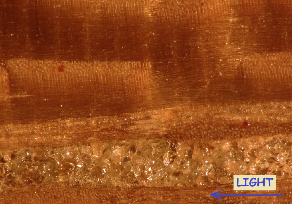
Sapele (pictures representing a 15x11mm area):
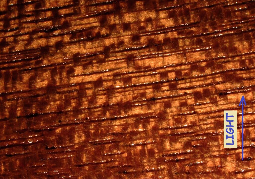
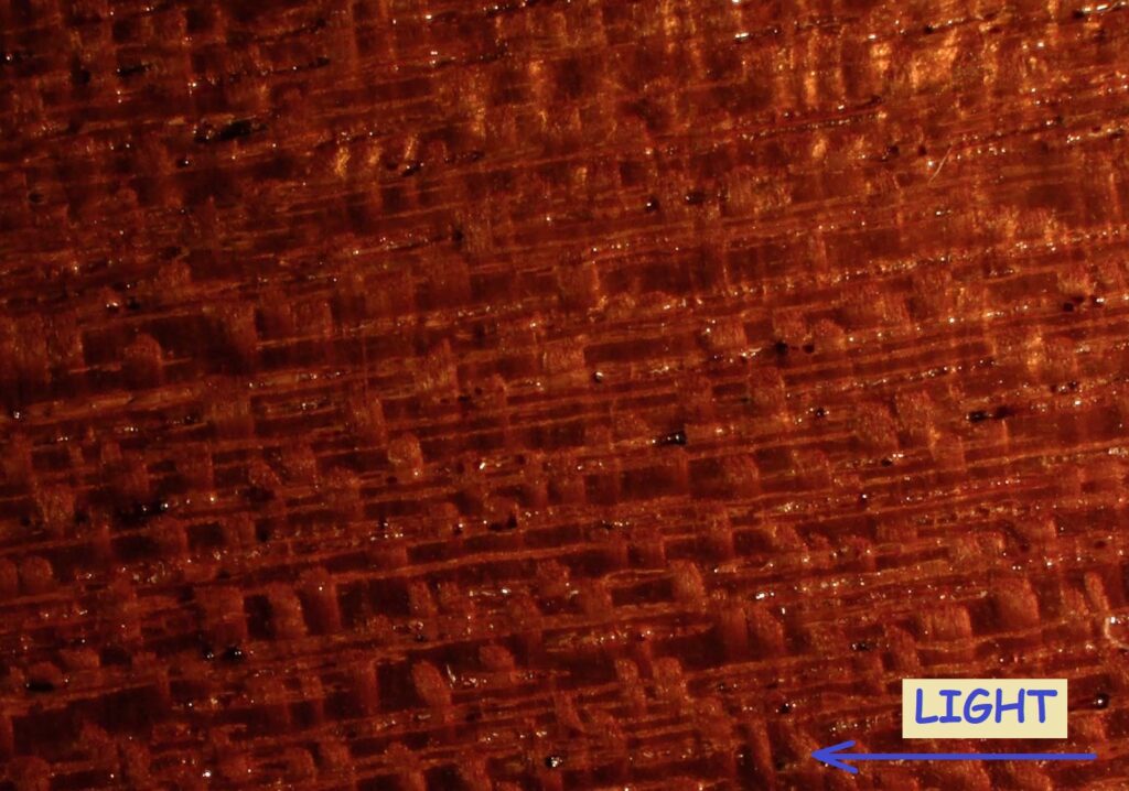
Sapele (pictures representing a 1.5×1.1mm area):
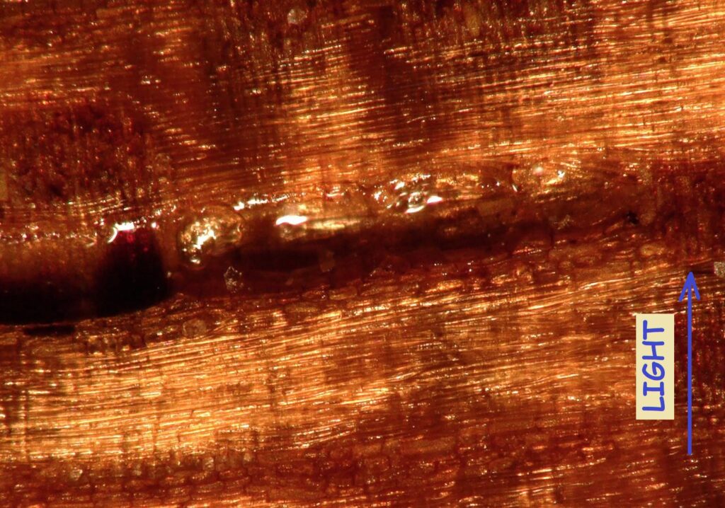
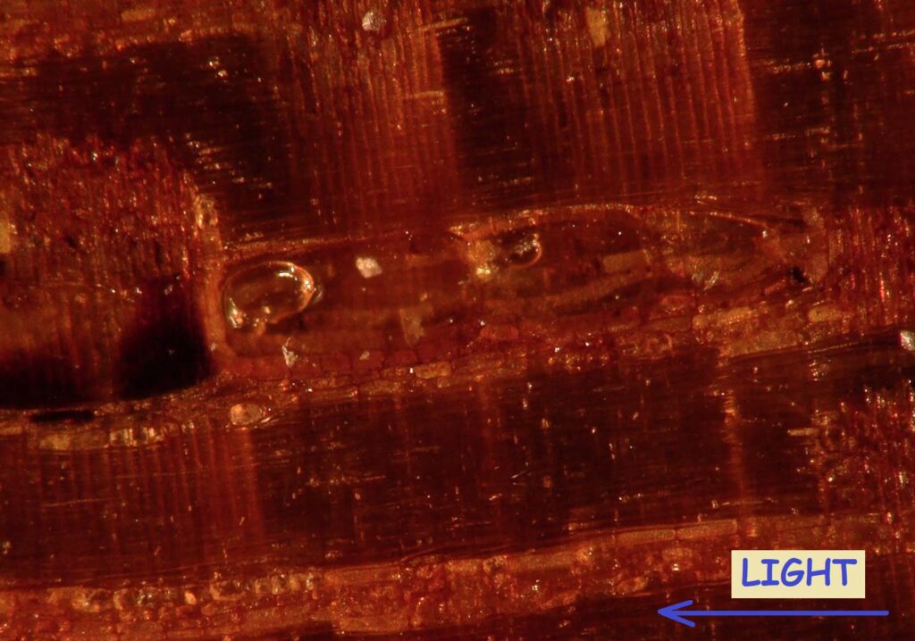
Black Walnut (pictures representing a 15x11mm area):
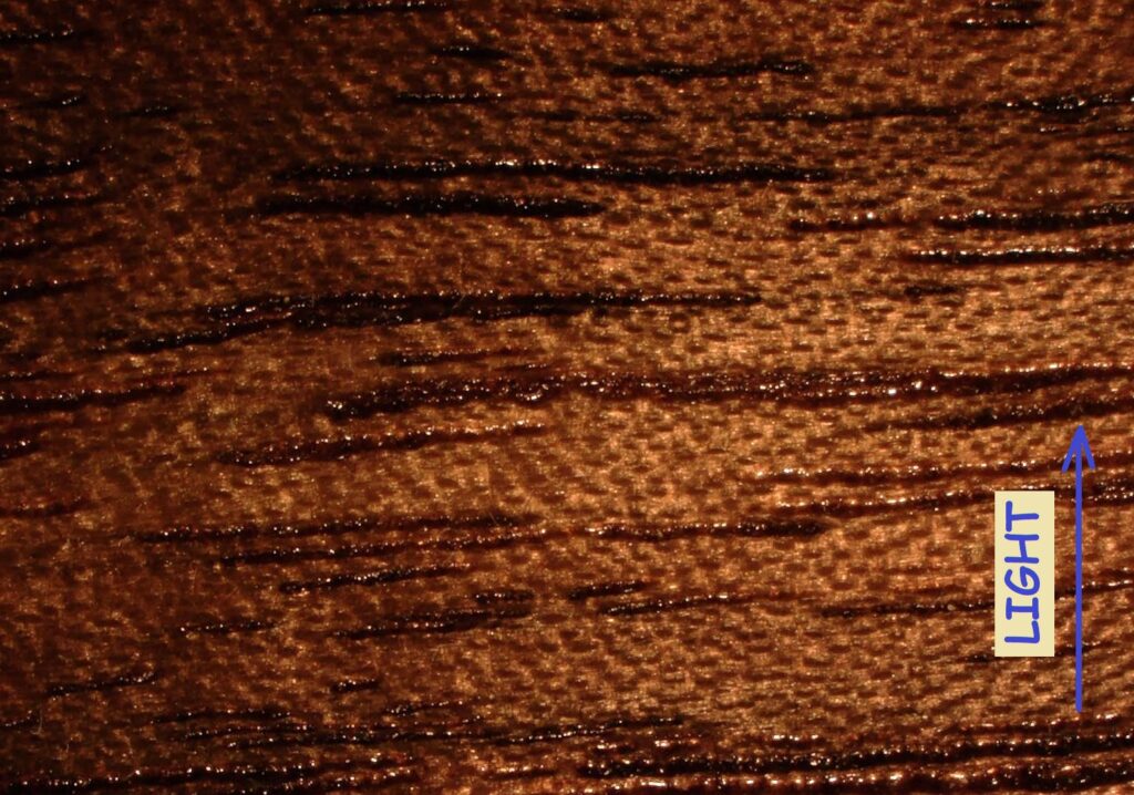
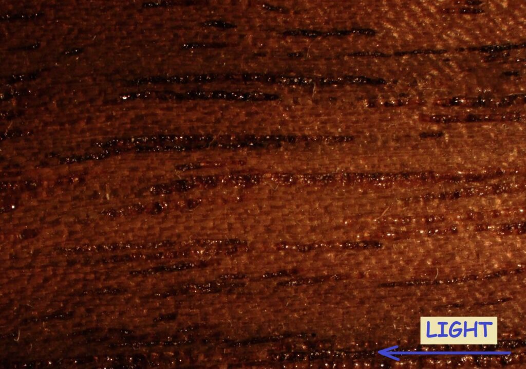
Black Walnut (pictures representing a 1.5×1.1mm area):
